Advanced Imaging Techniques in Houston: Understanding MRI, CT, and PET Scans
Imagine telling a doctor a century ago that those flickering silent screens would one day provide real-time pictures from inside a living person. They might have considered it impossible! One of the most amazing developments in modern medicine is our capacity to analyze the human body. You must be familiar with technologies like CT scans, MRIs, and PET scans. Maybe some people might have undergone these tests as well because being able to see more details inside the human body helps doctors in many ways. However, researchers within healthcare Houston are constantly striving for enhanced imaging techniques to innovate new technologies helping the disease diagnose faster for better health.
Advanced Imaging Techniques in Houston: Understanding MRI, CT, and PET Scans
MRI, CT, and PET scans have been proven to be much more than a miracle of life. But, do you really understand the difference between these imaging technologies? These scans offer detailed views of our organs and tissues, helping doctors diagnose and treat a wide range of conditions. It assists them to identify conditions ranging from minor issues like a fractured bone to serious diseases such as cancer or any brain disorders. This is why the doctors who specialize in these procedures are called radiologists. Many people don’t even realize that radiologists are the "real" doctors who play a crucial role in diagnosing the disease of patients behind the scenes.
MRI: Seeing Beyond the Bone
Magnetic resonance imaging (MRI) is a medical imaging technique that uses a combination of magnetic force and radio waves to create detailed pictures of organs, soft tissues, bones, and other internal structures. Unlike the technology of X-ray, which uses radiation, MRI does not involve any ionizing radiation.
While X-rays excel at imaging bones and detecting fractures, MRIs are better suited for examining soft tissues like muscles, ligaments, and the brain. It also makes MRI a valuable tool in professional sports for diagnosing injuries or muscle strains. Additionally, MRIs can be used alongside X-rays or CT scans to provide doctors with a more comprehensive picture of a patient's condition. In some cases, MRIs are more effective for identifying certain abnormalities, such as tumors or blood clots.
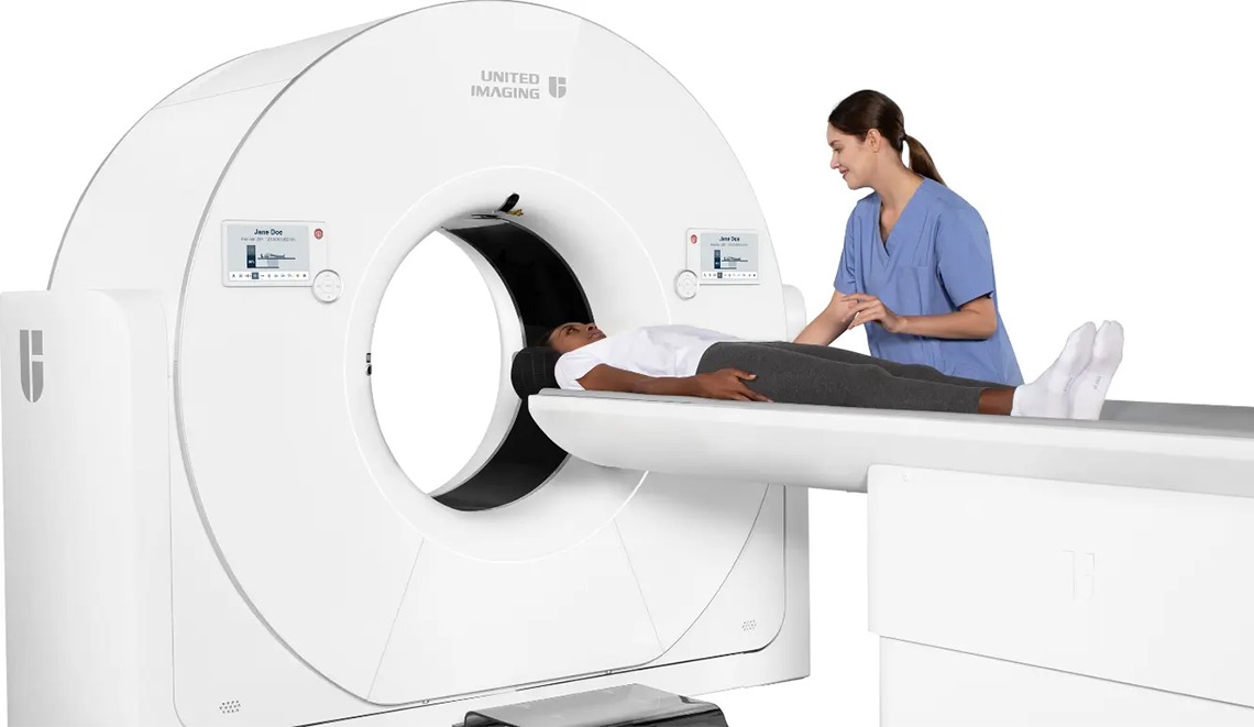
CT Scans: A 3D Perspective
CT scans, also known as computerized tomography scans, use X-ray technology. However, unlike a single X-ray, CT scans combine many X-rays taken from different angles and use advanced computer technology to provide much more detailed images.
The key advantage of CT scans lies in their ability to create detailed 3D images, unlike the flat 2D images produced by X-rays. This allows doctors to see a complete picture and identify even tiny details, making them highly useful for diagnosing and tracking internal bleeding, as well as pinpointing tumors and malignancies.
PET Scans: Viewing the Functions of Organs
Positron emission tomography, also known as a PET scan, is a powerful imaging technique. It uses tiny radioactive particles injected into your bloodstream to visualize your organs. These particles act like tracers, accumulating in areas with high blood flow, often indicating disease activity.
During a PET scan, a radiologist injects a safe amount of radioactive material into a vein. This material travels throughout your body, concentrating around organs with increased blood flow. A special scanner then detects the radiation emitted by the particles, creating detailed images of your organs. These images are transferred to a computer for analysis, allowing doctors to diagnose and monitor various conditions.
While not the first choice for initial cancer diagnosis, PET scans offer valuable insights for various conditions. They excel at revealing abnormalities in organs and blood flow, often indicating infections or inflammation. Additionally, PET scans provide a window into brain function, helping in diagnosing and monitoring diseases like Alzheimer's and epilepsy. However, PET scans provide less detailed anatomical information. To address this limitation, doctors often combine them with CT scans (PET-CT) for a more complete picture.
Some hospitals are now using a hybrid PET/MRI scan. This advanced technology produces highly detailed images with exceptional contrast. It is primarily employed by healthcare providers for diagnosing and monitoring cancers affecting delicate tissues such as the brain, head and neck, liver, and pelvis.
The Benefits of Modern Imaging
With advancements like MRI, CT and PET scans, doctors have even more powerful tools to diagnose and treat a wide range of conditions. It allows for more personalized care. These technologies not only optimize diagnostic capabilities but also contribute to safer and more effective patient care. As researchers in healthcare continue to innovate, the future holds promise for even more advanced imaging techniques that will further improve medical results while minimizing patient discomfort and exposure to radiation.